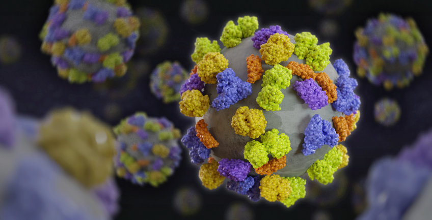
Nanoparticle Protein Interactions
By developing and combining different methods, we have excellently succeeded in establishing a worldwide unique characterization platform for the biomolecular corona of nanocarriers used as drug delivery vehicles. As soon as nanocarriers get in contact with biological fluids, proteins and other biomolecules like lipids and polysaccharides adsorb on the nanocarriers’ surface leading to a specific interaction behavior inside organisms.
Therefore, we have developed here at the institute a highly consistent workflow with label-free, quantitative proteomics based on cutting-edge liquid chromatography coupled to mass spectrometry (LC-MS) and combined this coherently with methods like differential scanning fluorimetry (NanoDSF), dynamic light scattering (DLS) in blood plasma, single protein investigations, 3D cryo electron microscopy and correlative light and electron microscopy (CLEM). Most notably this was done for the same nanocarriers over all platforms and also for situations which even extended to investigations in vivo or inside of cells.
Proteomics characterization of the biomolecular corona hereby has become a standard method in our characterizations for all biomedically interesting nanocarriers in our groups and has been extended to the intracellular protein corona for determining all steps during uptake, intracellular trafficking as well as exocytosis or degradation inside of cells. New preparation methods for obtaining the intracellularly trapped nanocarriers had to be developed. A highly resolved proteomic profile for each nanocarrier is the basis for targeted experiments further on with different proteins, e.g. apolipoproteins, antibodies, or complement factors. Also immunological functional cell assays for determining the ability of the nanocarriers to serve as nanovaccines have been realized and are used extensively for clinically applicable nanocarriers.
Differential scanning fluorimetry serves as a newly implemented tool to determine protein stability and/or conformation on nanocarrier surfaces. Via their intrinsic fluorescence, the thermal unfolding event of each protein can be monitored, so that protein denaturation can be detected. In contrast to other methods, nanoDSF can also be applied in the presence of nanocarriers (i.e. turbid dispersions) as long as no background fluorescence is present. Additionally, we apply multi-angle dynamic light scattering in undiluted blood plasma for each nanocarrier formulation that is developed for biomedical application as one of the few groups in the world. Using multi-exponential fitting procedures, aggregate formation can be detected very sensitively. This way, the highly important nanocarrier stability in blood after contact with proteins and other biomolecules can be assessed reliably. While even not accessible to many groups working with nanocarriers, both methods were implemented over the last years in our group to serve as standard quality control tools in the process of biomolecular corona characterization.
While nanoDSF can determine for bulk assembly of a given protein the denaturation, 3D cryo electron microscopy even more is suitable to explore the conformational changes of proteins on the nanocarrier surface. Here highly resolved structural changes down to near atomic resolution can be achieved. While this method is well established in the field of protein characterization, this method is actually developed in our group for questions of 3D structures of proteins in material science. It is based on single particle analysis and subtomogram averaging methods that have been successfully applied in structural biology, but needs to be transferred to the field of nanocarriers. Besides the unrivalled high resolution of the protein structure reconstruction, we expect this method to provide us with new insights into the interaction of proteins with surfaces. Due to the high resolution, we expect to be able to investigate how a protein is oriented towards the surface or interacts with the surface of the nanocarrier with a certain functional unit, e.g. a subdomain or secondary structure with the functional groups on nanocarriers.


