Unraveling Eumelanin Radical Formation by Nanodiamond Optical Relaxometry in a Living Cell
The generation of persistent, stable free radicals is a key feature of eumelanin physiochemistry. Scientists carried out EPR and IR to study the comproportionation reaction of eumelanin. The investigation of the formation of eumelanin radicals in the complex, dynamic and inhomogeneous environment of living cells has not yet been achieved, due to the lack of sensitivity of conventional detection methods. To gain a more comprehensive understanding of their biological functions in a spatiotemporal context, we developed a radical sensing and detecting nanodiamond (RGS-ND) quantum sensor that allows in situ real-time detection of the radicals present in eumelanin granules, with a particular emphasis on accurately quantifying the number of radicals formed inside cells.

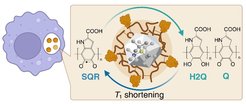
In our study, we first polymerized eumelanin on the surface of a nanodiamond quantum sensor. Due to the intrinsic sensitivity of the NV⁻ quantum sensor to magnetic field fluctuations, these NDs could serve as nanoscale sensors that are capable of quantitatively measuring the number of radical species in eumelanin. This was even possible at the level of individual cells, a regime inaccessible to standard EPR spectroscopy at such low radical levels. Combining the experimental T1 with theoretical simulations, we demonstrated that the number of radicals formed in the eumelanin layer is pH dependent. In comparison to EPR spectroscopy that provides valuable information on the presence and nature of paramagnetic species in various systems at the macroscopic level, we believe that T1 relaxometry could become an important tool for studying chemical reactions involving paramagnetic species in a nanoscale confined space that is not accessible by conventional techniques.
Using highly sensitive T1 relaxometry, we were able for the first time to monitor the chemical reaction in a layer of eumelanin just a few nanometers thick, even inside cells. We could quantify the number of radicals within the eumelanin shell even in a single living cell and we estimated the local pH value based on the number of detected radicals. It is the first time that radical species were detected in eumelanin with single cell resolution. Therefore, we believe that our method will shed light on the role of eumelanin in pigmentation, free radical scavenging, and antioxidation, which could also provide new insights into the melanin-related diseases to develop effective medical treatments.


