In a natural environment
Newly installed microscope offers unique examination method worldwide
 and Ingo Lieberwirth are looking forward for the first results using the newly installed Cryo-Electron microscope")
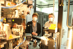
Katharina Landfester (left) and Ingo Lieberwirth are looking forward for the first results using the newly installed Cryo-Electron microscope
An electron microscope uses electrons to produce a magnified image of a sample. In doing so, it achieves an extremely high resolution, which makes it possible to image even the atoms in the examined sample. However, for this to work, the sample under investigation must meet certain requirements: For example, the sample must not change in the vacuum of the microscope. Water in particular, which is essential for the structural integrity of biological molecules, would evaporate immediately in the vacuum of the electron microscope, leading to drastic changes in the structure under investigation.
A new electron microscope has now been installed at the Max Planck Institute for Polymer Research in which the sample is shock-frozen at -190 °C - a so-called "cryo" electron microscope (from the Greek "kryos": cold). The result is that the water can no longer evaporate, allowing biological systems to be studied in their "natural" environment.
In addition, the microscope installed at MPI-P was combined with an energy spectrometer, making it unique in the world. The spectrometer measures the energy of the electrons and thus allows conclusions to be drawn about the atoms present in the sample. This enables the researchers at the MPI-P to measure the chemical composition of the molecules under investigation with almost atomic resolution.
The 5-million-euro microscope will complement the various microscopy techniques already available at the MPI-P and will contribute primarily to the structural elucidation of biomolecules. In particular, the new microscope will tackle previously unsolved questions at the boundary between molecular biology and materials science.
Video of the installation
"We are excited about the results we will be able to achieve with this microscope," says Ingo Lieberwirth, group leader and the person responsible for the microscope in Katharina Landfester's research group. "Especially in the field of using nanoparticles for medical applications, we will gain new insights into the mode of action of these nanoparticles to provide the basis for novel drugs."
The microscope will be officially inaugurated on June 25. The installation of the microscope has now been completed and work on its commissioning has already begun. The first measurements can therefore be expected in the next few weeks.


